Most people assume a dentist's before-and-after photos are just marketing. In reality, standardized clinical photography is one of the most rigorous diagnostic tools in cosmetic dentistry — and the rules behind it directly protect you as a patient. When a dentist follows an established photographic protocol, every smile they plan, every restoration they finish, and every case they publish has been documented under conditions designed to reveal the truth, not flatter it. Here is what that standard actually involves and why it should matter to anyone researching cosmetic dentistry in Clarksville, Tennessee. If you are here looking into cosmetic dental care, the sections below walk through the process from the first quote to the finished job.
What Is the 24-View Protocol?
The short version
- The AACD and ABCD require clinical case submissions in 24 standardized views: 12 before and 12 after under identical magnification, alignment, and lighting.
- The protocol calls for a RAW-capable DSLR with a manual-focus macro lens, and prohibits point-and-shoot cameras, softboxes, diffusers, and filters.
- Intraoral shots are taken at high f-stops of typically f/22 to f/32 so front and back teeth remain sharp in focus at the same time.
- Magnification ratios run 1:10 for the full face, 1:2 for full smile and parted arches, and 1:1 for anterior close-ups, converted for cropped sensors.
- At 1:1 magnification a single frame captures only four to six upper teeth, exposing a fraction of a millimeter of cement flash or a mismatched margin.
- The no-tilt rule requires the camera perpendicular at 90 degrees so a canted incisal plane or asymmetric gum line is photographed exactly as it exists.
The Equipment Rules Behind Every Frame
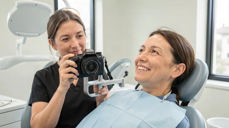
Standardized documentation starts with professional equipment. The protocol calls for a DSLR camera capable of shooting in RAW format — the uncompressed image file that preserves every detail the sensor captures — paired with a dedicated macro lens with manual focus and selectable magnification. Point-and-shoot cameras are not part of the standard. The light must be direct and undiffused, typically delivered by a lens-mounted ring flash or a dual-point macro flash; softboxes, diffusers, and filters are prohibited because they smooth over the very details the photographs exist to expose.
The operative field must be completely clean: no saliva film, no plaque, no pooled blood, no glove powder, no lipstick, no residual cement. An assistant typically manages the retractors, dries the teeth with an air syringe, and keeps the mirrors from fogging so every surface is visible. Intraoral shots are taken at high f-stops — typically f/22 to f/32 — so that front and back teeth remain in sharp focus at the same time.
Magnification: From 1:10 to 1:1
Each of the 24 views has a mandated magnification ratio, and those ratios are not arbitrary. They are calibrated so examiners can first evaluate the smile the way the human eye sees it, then zoom to microscopic levels where flaws become visible.
| View | Magnification | What It Reveals |
|---|---|---|
| Natural full face (frontal) | 1:10 | Facial symmetry, midline alignment, the smile within the face |
| Full smile (frontal and lateral) | 1:2 | Smile arc, gum display, buccal corridors, tooth proportions |
| Parted arches (frontal and lateral) | 1:2 | Incisal edges, embrasures, gingival contours and margins |
| Maxillary anterior close-ups (frontal and lateral) | 1:1 | Enamel micro-texture, translucency zones, restorative margins |
| Maxillary and mandibular arches (occlusal) | 1:2 | Full arch form, wear patterns, lingual and palatal surfaces |
At 1:1 magnification, a single frame captures only four to six upper teeth — and at that scale, a fraction of a millimeter of cement flash, a mismatched margin, or a dull patch of enamel texture is impossible to hide. For cameras with cropped sensors, the ratios are converted (roughly 1:15, 1:3, and 1:1.5 on the lens barrel) to keep the framing identical across different camera systems.
The No-Tilt Rule
One rule matters more than most patients realize: the camera must be held precisely perpendicular to the subject — 90 degrees both horizontally and vertically — and tilting it to compensate for crooked teeth, an uneven smile line, or asymmetric gums is a serious protocol violation. If a patient's incisal plane is canted, the photograph must show it. A dentist who refuses to tilt the camera is a dentist who refuses to hide your real anatomy from the record.
What Standardized Photos Mean for You
When you see a portfolio built on standardized documentation, you are looking at work that has survived brutal self-scrutiny. The dentist reviewed their own results at up to 10x magnification, found the flaws, and corrected them before you ever saw the finished case. When your own records are compared over time, standardized views let you and your dentist track changes in gum height, tooth wear, and margin integrity with genuine accuracy. It is the difference between marketing and medicine — and it is a standard worth looking for when you choose a cosmetic dentist in Clarksville, Tennessee.
If you are comparing providers in Clarksville, Tennessee, ask to see their clinical documentation the same way you would ask a surgeon for results. Look for retracted close-up views, consistent lighting between before and after shots, and no tilted camera angles. A dentist who photographs under the AACD standard has nothing to hide — and that transparency is exactly what you want protecting your smile.